G protein-coupled receptor (GPCR) – GCGR
02 Feb 2024
Share
Cryo-electron microscopy single-particle method is often used for structure determination of proteins which are difficult or impossible to crystallize. 2020, a structure information of GCGR protein which belongs to GPCR family was published in 《Science》, the structure determination of this protein was done by the experts now in ReadCrystal.
  • Background
    G protein-coupled receptors (GPCRs) belong to membrane proteins with extensive hydrophobic surfaces in the intramembrane region, which are difficult to stabilize in polar aqueous solutions after dissociation from the membrane and usually cannot use conventional crystallographic methods to obtain their structural information.
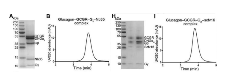
    The human glucagon receptor (GCGR), a member of the class B GPCR family, is essential for glucose homeostasis and is expected to be a potential drug target for the treatment of diabetes and obesity. In this 
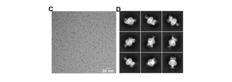
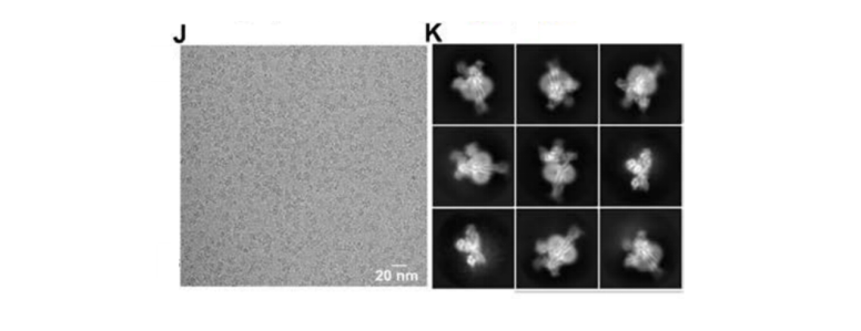
study, the structures of GCGR with its cognate ligand glucagon and heterotrimeric Gs or Gi1 protein were resolved by cryo-EM SPA to investigate the molecular mechanism of GCGR interaction with G proteins.
  • Results
    1. Protein expression & purification
    Expression of GCGR protein was extremely low, making it difficult to perform subsequent purification experiments. In order to obtain suitable protein complexes for further experiments, the vector and expression system were continuously adjusted. We also added monoclonal fragments to the protein expression process to increase the stability of the complexes.
    Fig. 1 Results of SDS-PAGE and analytical size-exclusion chromatography for the glucagon–GCGR–Gs–Nb35 complex (A, B) and glucagon–GCGR–Gi1–scFv16 complex (H, I)
  • 2. Sample preparation and Cryo-EM data acquisition
    During the protein preparation stage, the protein samples mostly stay on the grids’ support membrane rather than in the ice layer. Simultaneously, the preferred orientation of protein is serious.

    By adjusting the concentration of the protein and changing the grid type, the protein is eventually heavily oriented into the pores. The preferred orientation of the protein particles was improved by increasing the thickness of the frozen ice layer and laying a carbon membrane on the grid to average the number of particles with different orientations.
    Fig. 2 Cryo-EM image of Glucagon-GCGR-Gs-Nb35 complex
    Fig. 3 Cryo-EM image of Glucagon-GCGR-Gi1-scfv16 complex
  • 3. Structure determination
    The structural information of the glucagon-GCGR-G protein complex was obtained by data processing and 3D reconstruction.
    Fig. 4 Overall architectures of glucagon-GCGR–G protein complexes
    (A, B) Cryo-EM structure of the glucagon-GCGR-Gs-Nb35 complex at 3.7 Å resolution; (C, D) Cryo-EM structure of the glucagon-GCGR-Gi1-Scfv16 complex at 3.9 Å resolution
  • Publication
Related posts
Get in contact with us
  • +86 18962587269

  • contact@readcrystal.com

  • Changshu High-tech Industrial Development Zone, Suzhou, Jiangsu Province